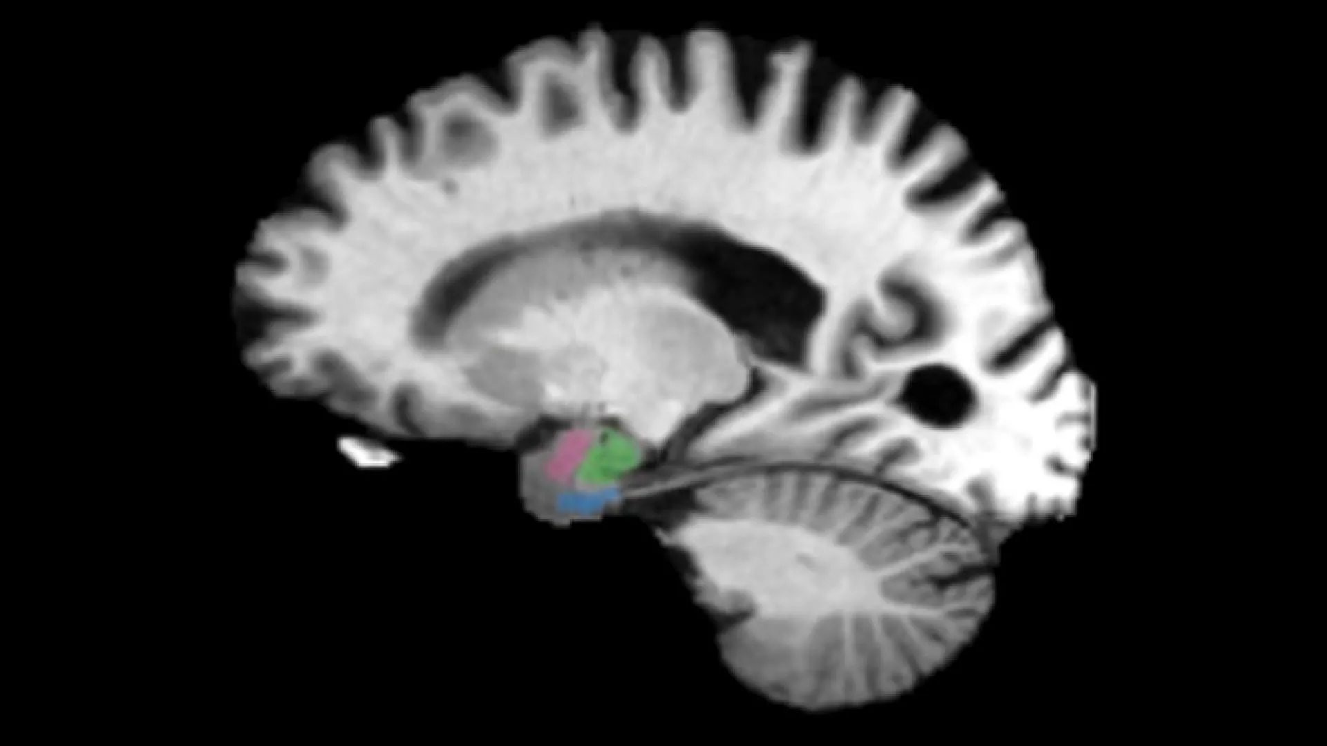
Alzheimer’s disease may produce detectable changes in two brain regions years before memory loss becomes apparent, according to researchers at Johns Hopkins University. Their analysis of hundreds of brain scans found earlier tissue loss in the entorhinal cortex and amygdala than in the hippocampus, the brain region commonly examined for signs of the disease.
The researchers said the changes could occur as much as a decade before clinical symptoms. They linked the earlier atrophy patterns to abnormal tau protein buildup, raising the prospect that imaging-based markers could help identify people at risk before substantial cognitive decline.
Looking beyond the hippocampus
The hippocampus has long been a focal point for assessing Alzheimer’s-related brain change because of its central role in memory. But the new work suggests it may not be the earliest region to show measurable deterioration.
The entorhinal cortex is involved in memory, while the amygdala is associated with emotion. In the Johns Hopkins analysis, both regions began losing tissue before the hippocampus. The researchers concluded that these regions predicted early Alzheimer’s disease better than the hippocampus in the scans they examined.
That difference could matter for the timing of detection. By the time memory problems prompt clinical attention, brain changes associated with Alzheimer’s may have been accumulating for years. Researchers have long understood that the disease develops over an extended pre-symptomatic period; the new findings focus attention on which anatomical signals may appear earliest in that process.

A long preclinical window
Senior author Michael Miller said the work identified clinical biomarkers of atrophy as much as a decade before clinical symptoms. He described a five-to-10-year period in which the brain may be changing before symptoms become apparent.
The possibility of a longer detection window is important because treatment strategies may be most useful before extensive neurodegeneration and cognitive impairment have occurred. The researchers said earlier recognition could eventually allow treatment to begin when it has a better chance of protecting the brain.
That is a potential future application, not an immediate diagnostic replacement. The supplied report describes the work as a finding from brain scans and says it could eventually help doctors recognize Alzheimer’s years before symptoms. It does not establish that the scan patterns alone can diagnose the disease in an individual person or predict with certainty who will develop it.
Tau and the pattern of atrophy
The study connects the early structural changes with abnormal tau protein buildup. Tau is one of the biological abnormalities associated with Alzheimer’s disease, and the report says that deterioration in the entorhinal cortex and amygdala was linked to it.
That connection gives the result relevance beyond an observation that some regions shrink earlier than others. It suggests that the sequence of atrophy may reflect underlying disease biology, potentially making regional brain changes useful alongside other biomarkers used in Alzheimer’s research and clinical assessment.
The finding also shifts the question from whether the hippocampus is important—it remains a key memory-related structure—to whether relying on it as the primary early anatomical signal leaves useful information untapped. In the Johns Hopkins data, the entorhinal cortex and amygdala offered earlier evidence of change.

What the research does and does not show
Alzheimer’s is the most common cause of dementia worldwide, but it does not begin when a person first notices forgetfulness or confusion. Disease-related changes can accumulate in the brain for decades before noticeable symptoms emerge. The research addresses a portion of that silent progression by identifying two regions that may be especially informative during the years before memory loss.
The report does not claim that routine screening should immediately shift to these regions, nor does it provide a treatment tested through this imaging approach. Instead, it adds evidence that the brain’s earliest measurable changes may be distributed differently from the later-stage signs that clinicians commonly associate with memory impairment.
Further work will be needed to establish how reliably these findings translate across people, imaging settings and stages of disease, and how they might be incorporated with other measures of Alzheimer’s risk and pathology. Still, the result offers a more specific target for researchers seeking markers of disease before symptoms emerge.
Research status
The findings were recently published in NeuroImage Reports and are being presented at Neuroscience 2026, the annual meeting of the Society for Neuroscience. The study’s central message is straightforward: signs of Alzheimer’s-related atrophy may appear in the entorhinal cortex and amygdala before the hippocampus and long before memory symptoms.
If confirmed and developed further, that pattern could help reshape how researchers look for the earliest observable effects of Alzheimer’s disease—and how early they may eventually be able to intervene.
This article is based on reporting by Science Daily. Read the original article.

Originally published on sciencedaily.com