लंडन पथकाने पहिल्या यशस्वी एआय-सहाय्यित मेंदूतील गाठ काढण्याची नोंद केली
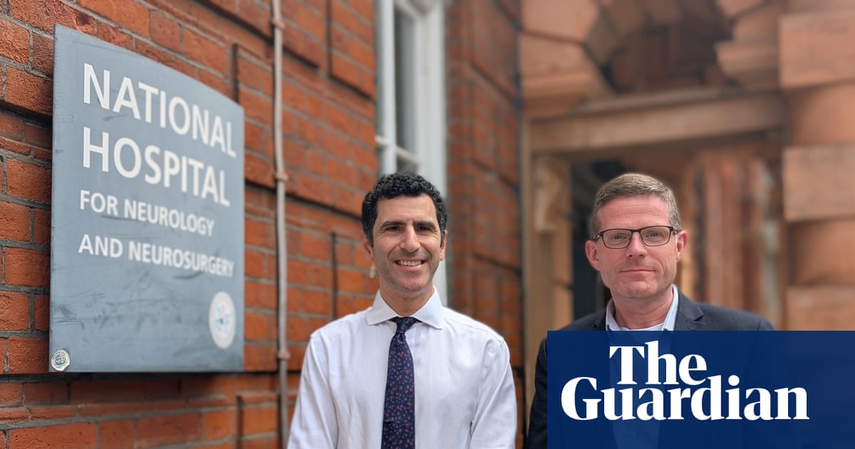
लंडनमधील National Hospital for Neurology and Neurosurgery येथील न्यूरोसर्जनांनी आरोग्य अधिकाऱ्यांच्या मते जगातील पहिली यशस्वी एआय-सहाय्यित मेंदूतील गाठ काढण्याची शस्त्रक्रिया केली. मे महिन्यात करण्यात आलेल्या आणि रुग्ण बरा झाल्यानंतर उघड करण्यात आलेल्या या प्रक्रियेत शस्त्रक्रियेच्या वेळी थेट कॅमेरा फुटेजचे विश्लेषण करण्यासाठी कृत्रिम बुद्धिमत्ता प्रणाली वापरण्यात आली, ज्यामुळे गाठीच्या परिसरात अतिशय जवळ असलेल्या संवेदनशील रचनांची ओळख पटवता आली.
४८ वर्षीय Rhys Hibbert यांना पिट्यूटरी ग्रंथीमध्ये ११-मिलीमीटरची गाठ होती, अशा भागात जिथे दृष्टी नियंत्रित करणाऱ्या रक्तवाहिन्या आणि नसांचा परस्परांशी अतिशय जवळचा संबंध असतो. दिलेल्या स्रोत मजकुरानुसार, शस्त्रक्रिया पथक संपूर्ण ऑपरेशनदरम्यान पूर्ण नियंत्रणात होते, तर एआय प्रणालीने टाळावयाच्या नसां आणि रक्तवाहिन्यांसारख्या महत्त्वाच्या शरीररचनांना अधोरेखित केले.
नोंदवलेला परिणाम या प्रकरणाला केवळ एका रुग्णालयाच्या यशापेक्षा अधिक महत्त्व देतो. अत्यंत मर्यादित मेंदूविषयक प्रक्रियांमध्ये, शल्यचिकित्सक अनेकदा अशा जागेत काम करतात जिथे एका मिलीमीटरची चूकही अंधत्व, स्ट्रोक किंवा मृत्यू यांसारख्या गंभीर परिणामांना कारणीभूत ठरू शकते. चिकित्सकांकडून नियंत्रण न घेता प्रत्यक्ष वेळेत शरीररचनेची ओळख सुधारू शकणारे साधन ऑपरेशन थिएटरमध्ये एक महत्त्वाचा सहाय्यक थर ठरू शकते.
एआय प्रणालीचा वापर कसा करण्यात आला
शस्त्रक्रियेच्या वेळी थेट एंडोस्कोपिक कॅमेरा प्रतिमांचे विश्लेषण करून आणि शल्यचिकित्सकांना जलद व सुरक्षितपणे ओळखणे आवश्यक असलेल्या रचनांना रंग-कोड करून ही प्रणाली कार्य करत होती. पिट्यूटरी शस्त्रक्रियेमध्ये हे विशेषतः महत्त्वाचे आहे, कारण तेथील शरीररचना गर्दीची आणि चूक न माफ करणारी असते. पिट्यूटरी ग्रंथी दृष्टी आणि रक्तप्रवाहासाठी आवश्यक असलेल्या रचनांच्या जवळ असते, आणि ऑपरेटिव्ह परिस्थितीत होणारा कोणताही गोंधळ त्वरित परिणाम देऊ शकतो.
स्रोत मजकुरात University College London येथील robotics and AI मधील associate professor आणि प्रणालीसाठी technical lead म्हणून ओळखल्या गेलेल्या डॉ. Sophia Bano यांनी सांगितले की मॉडेलला शेकडो शस्त्रक्रिया व्हिडिओंवर प्रशिक्षित करण्यात आले होते. हे प्रशिक्षण तंत्रज्ञानाच्या मुख्य ताकदींपैकी एक म्हणून मांडले जाते: त्यामुळे सॉफ्टवेअरला अशा अनेक मागील शस्त्रक्रिया उदाहरणांमधून शिकता येते, जी एका चिकित्सकाला प्रत्यक्ष अनुभवातून मिळवण्यासाठी वर्षे लागू शकतात.
व्यावहारिकदृष्ट्या, एआयने शस्त्रक्रियेच्या निर्णयक्षमतेची जागा घेतली नाही. त्याऐवजी, ते संदर्भात्मक इशारा आणि ओळख साधन म्हणून कार्य करत होते, ज्यामुळे पथकाला ऊतकांचे प्रकार वेगळे ओळखता आले आणि हात लावू नयेत अशा रचनांचा शोध घेता आला. हा फरक महत्त्वाचा आहे. वर्णन केल्याप्रमाणे, हे प्रकरण स्वयंचलित शस्त्रक्रियेबद्दल नाही. हे augmentation बद्दल आहे: सॉफ्टवेअर जे शल्यचिकित्सक आधीच पाहत आहेत त्याची दृश्यमानता आणि अर्थ लावण्याची क्षमता वाढवते.

हे प्रकरण वेगळे का ठरते
रुग्णालये आणि संशोधन गट अनेक वर्षांपासून radiology, pathology, workflow optimization, आणि surgical planning मध्ये एआय प्रणालींची चाचणी करत आहेत. ही बातमी उल्लेखनीय ठरण्याचे कारण म्हणजे ही तंत्रज्ञान एखाद्या जिवंत रुग्णाच्या प्रक्रियेत वापरण्यात आली, मागोवा घेणाऱ्या संशोधन साधनाप्रमाणे नव्हे. स्रोत मजकुरात म्हटले आहे की रुग्णालयाने याआधी ही प्रणाली संशोधनासाठी वापरली होती, पण प्रत्यक्ष शस्त्रक्रियेत ती प्रथमच वापरली गेली.
संशोधन वातावरणातून रुग्णसेवेकडे जाणे हे अनेक वैद्यकीय एआय कार्यक्रमांसाठी सर्वात कठीण आव्हान असते. विकास वातावरणातील कामगिरी थेट सुरक्षित क्लिनिकल वापरात आपोआप रूपांतरित होत नाही, विशेषतः शस्त्रक्रियेदरम्यान, जिथे वेळेची मर्यादा तातडीची असते आणि अपयशासाठी सहिष्णुता जवळपास शून्य असते. म्हणूनच यशस्वी पहिल्या प्रकरणाला प्रतीकात्मक आणि कार्यात्मक अशा दोन्ही पातळ्यांवर वजन आहे: त्यातून दिसते की पथकाच्या मते प्रणाली प्रत्यक्ष परिस्थितीत मदत करण्याइतकी परिपक्व झाली होती.
रुग्णाच्या बरे होण्यामुळे या घोषणेचा प्रभाव अधिक ठळक झाला. Hibbert म्हणाले की ते जागे झाल्यावर त्यांना खोलीतील वस्तू स्पष्ट दिसत होत्या. दिलेल्या स्रोत साहित्याप्रमाणे, आठवड्याभरात ते चष्म्यांशिवाय किंवा काठीशिवाय स्वतंत्रपणे चालू शकले, आणि नंतर ते पुन्हा कामावर परतले. हे तपशील वैद्यकीय दावे प्रत्यक्षात उतरवतात. ही केवळ सोयीतील किरकोळ सुधारणा नव्हती. ही शस्त्रक्रिया थेट कार्यक्षमतेचे आणि जीवनाच्या गुणवत्तेचे संरक्षण करण्याशी संबंधित होती.
शस्त्रक्रियेसाठी याचा काय अर्थ असू शकतो
भविष्यातील प्रकरणांनीही तोच परिणाम दर्शवला, तर अशा प्रकारची एआय मदत विशेषतः अशा शस्त्रक्रियांमध्ये उपयुक्त ठरू शकते जिथे लहान संरचना जलद आणि विश्वासार्हपणे ओळखाव्या लागतात. न्यूरोसर्जरी हा स्पष्ट पर्याय आहे, कारण त्यात अचूकतेची अत्यंत गरज असते, पण अशाच image-guided मदतीचा उपयोग इतर minimally invasive क्षेत्रांमध्येही होऊ शकतो, जिथे शरीररचना दृश्यदृष्ट्या गुंतागुंतीची आणि साधनांचा प्रवेश मर्यादित असतो.
मोठा अर्थ असा की शस्त्रक्रियात्मक एआय कदाचित प्रथम अरुंद, काळजीपूर्वक मर्यादित वापरांद्वारे पुढे जाईल, व्यापक automation द्वारे नव्हे. real-time annotation, risk highlighting, आणि शरीररचना ओळख ही प्रक्रियेचे नियंत्रण सॉफ्टवेअरकडे सोपवण्यापेक्षा क्लिनिकलदृष्ट्या अधिक सहजपणे समर्थनयोग्य आहेत. त्या आधुनिक शस्त्रक्रियेच्या पद्धतीशी अधिक नैसर्गिकरीत्या जुळतात, जिथे चिकित्सक navigation systems, imaging, आणि robotics एका स्तरित निर्णयप्रक्रियेत एकत्र करतात.
यामुळे कठीण प्रश्न नाहीसे होत नाहीत. नोंदवलेल्या शस्त्रक्रियेच्या फुटेजवर प्रशिक्षित प्रणालींनी हे सिद्ध करावे लागेल की त्या विविध शरीररचना, प्रकाश, कॅमेरा कोन आणि दुर्मीळ सादरीकरणांमध्येही विश्वासार्ह राहतात. सॉफ्टवेअरचे आउटपुट स्पष्ट, जलद आणि सुसंगत असावेत, जेणेकरून ते विचलित न करता मदत करतील, याची खात्री रुग्णालयांना हवी आहे. स्रोत मजकूर या समस्या सुटल्या असल्याचा दावा करत नाही, पण या प्रकरणातून असे दिसते की एक पथक सिद्धांतापलीकडे जाऊन काळजीपूर्वक व्यवस्थापित प्रत्यक्ष वापरात आले आहे.

एक मैलाचा दगड, पूर्ण झालेली कहाणी नाही
लंडनमधील ही प्रक्रिया क्लिनिकल एआयमधील एक सुरुवातीचा मैलाचा दगड म्हणून समजली पाहिजे, विकास चक्राचा शेवट म्हणून नव्हे. एक यशस्वी शस्त्रक्रिया स्वतःहून व्यापक परिणामकारकता सिद्ध करत नाही. पण निवडक प्रकरणांमध्ये शस्त्रक्रियेत एआय मदत संकल्पना-प्रदर्शनांमधून पुढील टप्प्यात, म्हणजे अग्रभागी रुग्णसेवेकडे, जात असल्याचे ती दाखवते.
आरोग्य व्यवस्था, संशोधक आणि वैद्यकीय उपकरण विकासकांसाठी हा बदलच खरा विषय आहे. तांत्रिक प्रश्न आता फक्त एआय शस्त्रक्रियेच्या व्हिडिओमध्ये रचना ओळखू शकते का, इतकाच राहिलेला नाही. शल्यचिकित्सक मानवी अचूकतेच्या टोकावर काम करत असताना, अशी ओळख प्रत्यक्ष वेळेत, सुरक्षित, विश्वासार्ह आणि खरोखर उपयुक्त पद्धतीने देता येते का, हा खरा प्रश्न आहे.
या प्रकरणात उत्तर होकारार्थी वाटते. प्रणालीने एका उच्च-जोखीम ऑपरेशनमध्ये शल्यचिकित्सा पथकाला मार्गदर्शन करण्यात मदत केली, आणि रुग्णाची दृष्टी जपली गेली. तांत्रिक मदत आणि मानवी नियंत्रण यांचे हे संयोजन मशीन-प्रतिस्थापनाच्या rhetoric पेक्षा वैद्यकीय एआय स्वीकाराच्या पुढच्या टप्प्याला अधिक परिभाषित करेल अशी शक्यता आहे. सध्या तरी, लंडनमधील ही नोंदवलेली पहिली घटना ऑपरेशन थिएटरमध्ये त्या भविष्यातील एक ठोस उदाहरण देते.
हा लेख The Guardian च्या वार्तांकनावर आधारित आहे. मूळ लेख वाचा.
Originally published on theguardian.com