Introduction: A New Paradigm in Neuroimaging AI
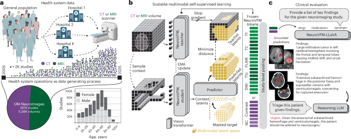
Artificial intelligence has shown remarkable promise in medical imaging, but most models are narrow—trained to detect a single disease using curated datasets. A new study published in Nature Medicine introduces NeuroVFM, a generalist neuroimaging model trained on routine clinical MRI and CT scans from health systems. By leveraging health system learning, NeuroVFM captures broad, generalizable representations of brain anatomy and pathology, outperforming task-specific models across multiple diagnostic scenarios.
What Is NeuroVFM?
NeuroVFM stands for Neuroimaging Vision Foundation Model. Unlike traditional models that are trained from scratch for each task, NeuroVFM is pre-trained on a large, diverse corpus of real-world clinical scans—including both MRI and CT modalities—collected from routine care. This approach allows the model to learn fundamental features of brain structure and common abnormalities without manual annotation. The researchers used a self-supervised learning technique, enabling the model to learn from unlabeled data by predicting missing parts of images or contrasting different views.

Health System Learning: Why It Matters
Most medical AI models are trained on high-quality, curated datasets that may not reflect real-world variability. In contrast, NeuroVFM was trained on scans from multiple health systems, encompassing a wide range of scanner manufacturers, protocols, patient demographics, and pathological conditions. This diversity makes the model robust to domain shifts—a common challenge when deploying AI in new hospitals. The study shows that NeuroVFM's representations generalize better than those from models trained on smaller, cleaner datasets.
Performance Across Multiple Tasks
The researchers evaluated NeuroVFM on several downstream tasks, including brain tumor segmentation, intracranial hemorrhage detection, and Alzheimer's disease classification. In each case, NeuroVFM either matched or exceeded the performance of state-of-the-art task-specific models. For example, in tumor segmentation, NeuroVFM achieved Dice scores comparable to dedicated models while requiring fewer labeled examples for fine-tuning. In hemorrhage detection, it showed higher sensitivity and specificity across different CT scanner types.

Implications for Clinical Practice
NeuroVFM's generalist nature could streamline clinical workflows. Instead of deploying multiple AI tools for different conditions, hospitals could use a single model that handles a variety of neuroimaging tasks. This reduces computational overhead and simplifies maintenance. Moreover, because NeuroVFM learns from routine scans, it can be continuously updated with new data, adapting to evolving clinical practices and emerging diseases.
Limitations and Future Directions
While promising, NeuroVFM has limitations. The study did not include all rare neurological conditions, and the model's performance on extremely low-resolution or artifact-heavy scans needs further validation. Additionally, the self-supervised pre-training requires substantial computational resources. Future work may explore more efficient training methods and expand the model to include other imaging modalities like PET or functional MRI.

Conclusion
NeuroVFM represents a significant step toward generalist AI in neuroimaging. By harnessing health system learning, it achieves robust, generalizable representations that can improve diagnostic accuracy and efficiency. As healthcare AI moves toward foundation models, NeuroVFM offers a blueprint for building versatile tools that learn from the rich, messy data of real-world clinical practice.
This article is based on reporting by Nature Medicine. Read the original article.
Originally published on nature.com