Introduction
Aging is a complex biological process that manifests differently across tissues and organs. While chronological age is a universal metric, biological age varies significantly between individuals and even between organs within the same person. This variability has profound implications for healthspan and disease susceptibility. Now, a groundbreaking study published in Nature Medicine harnesses the power of whole-slide histopathological images to uncover morphological changes associated with aging across 40 tissue types. These 'histological aging signatures' promise to refine our understanding of tissue-specific aging and its link to disease.
The Study: A Pan-Tissue Analysis
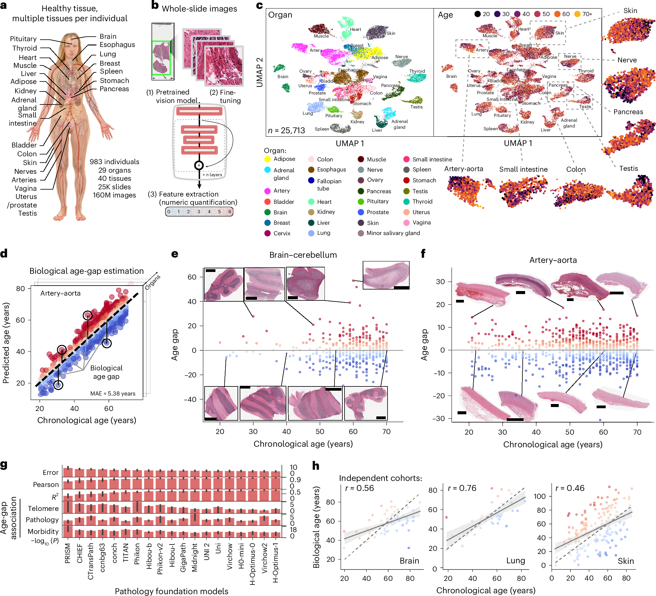
Researchers analyzed a vast collection of whole-slide histopathological images from 40 distinct tissue types, sourced from large-scale repositories. Using advanced computational techniques, they identified reproducible morphological features that correlate with chronological age. These features include changes in cellular density, nuclear morphology, tissue architecture, and the presence of age-related markers such as fibrosis or atrophy. The study's scale and breadth are unprecedented, providing a comprehensive atlas of aging across the human body.
Key Findings
The analysis revealed that each tissue exhibits a unique aging trajectory, with some tissues aging faster than others. For instance, certain tissues showed pronounced morphological changes early in life, while others remained relatively stable until later decades. Importantly, the researchers found that deviations from the expected aging trajectory—where a tissue appears 'older' or 'younger' than the individual's chronological age—were associated with specific diseases. This suggests that histological aging signatures could serve as biomarkers for disease risk and progression.

Implications for Disease Monitoring
The ability to assess biological age at the tissue level opens new avenues for disease monitoring. For example, a tissue that appears prematurely aged might be at higher risk for cancer, fibrosis, or degenerative conditions. Conversely, tissues that appear younger than expected might indicate resilience or protective factors. By integrating these signatures into clinical practice, physicians could potentially identify at-risk individuals earlier and tailor preventive strategies accordingly.
Potential Clinical Applications
One of the most promising applications is in oncology. Tumor-adjacent normal tissue often shows signs of accelerated aging, which could influence cancer progression and treatment response. Histological aging signatures might help stratify patients based on their tissue-level biological age, guiding decisions about surgery, chemotherapy, or immunotherapy. Similarly, in chronic diseases like kidney disease or liver cirrhosis, these signatures could track disease progression and response to therapy more accurately than current biomarkers.
Methodological Innovations
The study leverages state-of-the-art machine learning algorithms to extract and quantify morphological features from histopathology slides. This approach overcomes the limitations of manual assessment, which is subjective and time-consuming. The algorithms were trained on a diverse dataset and validated across multiple cohorts, ensuring robustness and generalizability. The resulting signatures are not only reproducible but also interpretable, as they correspond to known biological processes such as cellular senescence, inflammation, and extracellular matrix remodeling.
Data and Reproducibility
All data and code used in the study have been made publicly available, fostering transparency and collaboration. This open-science approach will enable other researchers to validate the findings in independent cohorts and explore new applications. The authors also provide detailed documentation, making it easier for the scientific community to adopt these methods.

Broader Implications for Aging Research
This study contributes to a growing body of evidence that aging is not a uniform process but rather a mosaic of tissue-specific changes. The concept of 'aging clocks' has gained traction in recent years, with epigenetic and proteomic clocks offering insights into biological age. Histological aging signatures add another dimension by capturing structural changes that are directly visible in tissue samples. This multimodal approach could lead to a more holistic understanding of aging and its heterogeneity.
Future Directions
The researchers plan to expand their analysis to include more tissue types and longitudinal samples, which would allow them to track aging trajectories over time. They also aim to integrate histological signatures with other omics data, such as transcriptomics and proteomics, to uncover the molecular mechanisms underlying morphological changes. Ultimately, the goal is to develop a comprehensive aging atlas that can be used to predict health outcomes and guide interventions.
Challenges and Considerations
While the findings are promising, several challenges remain. The study relies on histopathological images, which are typically obtained from biopsies or surgical specimens. This limits the applicability to tissues that are routinely sampled. Additionally, the association between histological aging and disease is correlational, and causal relationships need to be established through functional studies. Ethical considerations also arise regarding the use of aging biomarkers in clinical decision-making, particularly in the context of insurance or employment.
Validation and Standardization
For these signatures to be clinically useful, they must be validated in diverse populations and standardized across laboratories. The current study provides a strong foundation, but further work is needed to ensure that the algorithms perform consistently across different staining protocols, scanners, and image resolutions. Collaborative efforts will be essential to establish guidelines for clinical implementation.

Conclusion
The identification of histological aging signatures represents a significant step forward in precision medicine. By providing a detailed view of how aging manifests in different tissues, this research offers new tools for disease monitoring and risk assessment. As the field moves toward integrating these signatures into routine practice, we can anticipate a future where aging is managed proactively, and diseases are detected and treated earlier. The study not only advances our scientific understanding but also paves the way for personalized healthcare that accounts for the unique aging patterns of each individual.
This article is based on reporting by Nature Medicine. Read the original article.
Originally published on nature.com