How Blood Flow Follows Brain Activity
For over a century, scientists have understood that when a region of the brain becomes active, blood flow to that region increases to deliver oxygen and nutrients. This phenomenon, known as neurovascular coupling, underpins many modern brain imaging techniques, including functional magnetic resonance imaging (fMRI), which detects changes in blood oxygenation to map neural activity. However, the precise relationship between the type of neural activity and the vascular response has remained less clear. Are all neural activations treated equally, or does the brain tailor its blood supply to the specific demands of different sensory modalities?
A new study published in Science sheds light on this question. The research, titled "Modality-specific neurovascular coupling via layer-segregated arteriole networks," suggests that the brain's blood flow response is not a generic reaction but is precisely tuned to the type of sensory information being processed. The study indicates that different sensory modalities—such as vision, touch, or hearing—activate distinct vascular pathways in the cerebral cortex, and that these pathways are organized according to the layered structure of the cortex.
What Is Neurovascular Coupling?
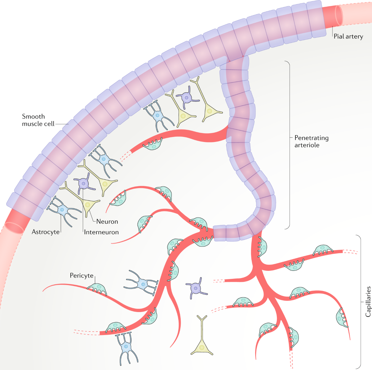
Neurovascular coupling is the intricate communication system between neurons and blood vessels. When neurons fire, they release chemical signals that cause nearby arterioles to dilate, increasing blood flow to the active area. This ensures that the brain's most active regions receive a disproportionate share of oxygen and glucose. The process is essential for normal brain function, and its breakdown is implicated in a range of disorders, including stroke, dementia, and hypertension.
Traditionally, neurovascular coupling has been viewed as a relatively uniform mechanism: active neurons in any part of the brain trigger a local increase in blood flow. But the new study challenges this notion by proposing that the coupling is modality-specific—meaning that the vascular response is tailored to the sensory system that is engaging the cortex.

Layer-Segregated Arteriole Networks
The cerebral cortex is arranged in layers, each with distinct cell types and connectivity patterns. Different sensory inputs target specific layers. For example, in the primary somatosensory cortex, inputs from the skin arrive in layer 4, while inputs from higher-order areas may target layers 2 and 3. The new research suggests that arterioles—the small blood vessels that control blood flow—are similarly segregated by cortical layer. Each layer has its own arteriole network, and these networks respond selectively to the neural activity originating in that layer.
According to the study's title, this layer-segregated organization allows for modality-specific blood flow regulation. When a particular sensory modality is stimulated, it activates the neural populations in specific cortical layers, which in turn communicates with the arterioles in those layers. This results in a precise, localized increase in blood flow that matches the exact neural circuits being used.
Implications for Brain Imaging
The findings have potentially significant implications for neuroimaging. fMRI measures blood flow and oxygenation changes as a proxy for neural activity. If the vascular response is modality-specific and layer-segregated, then fMRI signals could be interpreted more finely. Currently, standard fMRI has limited spatial resolution and often cannot distinguish between activity in different cortical layers. But if researchers can develop imaging techniques that exploit the layer-specific vascular responses, they might be able to infer not just which brain region is active, but which neural circuits within that region are engaged.
The study may also help explain why some fMRI studies have found conflicting results when comparing different sensory tasks. If different tasks elicit different vascular responses due to their laminar profiles, then comparing raw BOLD signals across tasks could be misleading. A deeper understanding of modality-specific neurovascular coupling could lead to more accurate calibration of fMRI signals, making them a truer reflection of underlying neural activity.

Potential Clinical Significance
Beyond brain imaging, the research could inform our understanding of neurological and psychiatric disorders. Many conditions, including Alzheimer's disease, schizophrenia, and migraine, are associated with disruptions in neurovascular coupling. If the coupling is modality-specific, then these disruptions might not affect all sensory systems equally. For example, a patient with a vascular pathology might show preserved blood flow for some sensory tasks but impaired responses to others, depending on which cortical layers and arteriole networks are involved. This could lead to new diagnostic approaches that use targeted sensory stimulation to test the integrity of specific vascular pathways.
Furthermore, the study's insights into layer-specific vascular architecture could inspire new strategies for drug delivery to the brain. If arteriole networks are segregated by layer, then drugs that modulate vascular tone could be designed to target specific networks, potentially enhancing delivery of therapeutics to particular cortical areas while minimizing side effects elsewhere.
Methodological Considerations
While the study's title provides a clear summary, the full details of the experimental methods and findings are not yet publicly available. However, based on the title, it is evident that the researchers employed techniques capable of measuring both neural activity and vascular responses with high temporal and spatial resolution, likely in animal models. Advances in two-photon microscopy and optogenetics have made it possible to observe neural activity and blood vessel diameter simultaneously in vivo, giving scientists an unprecedented look at the subtle dynamics of neurovascular coupling.
It is also worth noting that the study was published in the prestigious journal Science, which indicates that the findings are likely to be rigorous and have broad implications for the field.
Key Takeaways
- Neurovascular coupling may be modality-specific, with different sensory systems driving distinct blood flow responses.
- These responses are mediated by arteriole networks that are segregated according to cortical layer.
- The findings could improve fMRI interpretation by enabling layer-specific imaging of brain activity.
- Disruptions in modality-specific coupling may have clinical relevance for diseases affecting the brain's blood supply.
- The study opens new avenues for targeted therapeutic interventions based on vascular architecture.

Future Directions
The study likely marks the beginning of a deeper exploration into the relationship between brain architecture and blood flow. Future research will need to confirm whether these layer-segregated networks are conserved across species and whether they exist in all regions of the cortex. Additionally, researchers will investigate how these networks are modulated by age, disease, and other factors. The ultimate goal is to understand how the brain's vascular system is finely tuned to support its complex computational tasks.
As we move forward, this research reminds us that the brain's support systems are as sophisticated as the neurons themselves. The vascular network is not merely a plumbing system but an integrated component of brain function, dynamically adapting to the brain's ever-changing needs. By uncovering the modality-specific and layer-segregated nature of neurovascular coupling, the study provides a crucial piece of the puzzle in understanding how the brain allocates its resources.
In conclusion, the study offers a fresh perspective on the brain's blood flow regulation, suggesting that sensory information is processed with remarkable vascular precision. This work not only enhances our fundamental understanding of cerebral physiology but also holds promise for improving diagnostic and therapeutic approaches in neurology.
This article is based on reporting by Science (AAAS). Read the original article.
Originally published on science.org