A new lens on how men and women age
A paper published in Nature Medicine on 16 September 2026 introduces what the journal's summary describes as sex-specific biological aging clocks spanning multiple organs and molecular systems (doi:10.1038/s41591-026-04662-6). The central premise is straightforward but consequential: the biological processes that accompany aging do not unfold identically in female and male bodies, and the instruments used to measure those processes may need to reflect that difference rather than average it away.
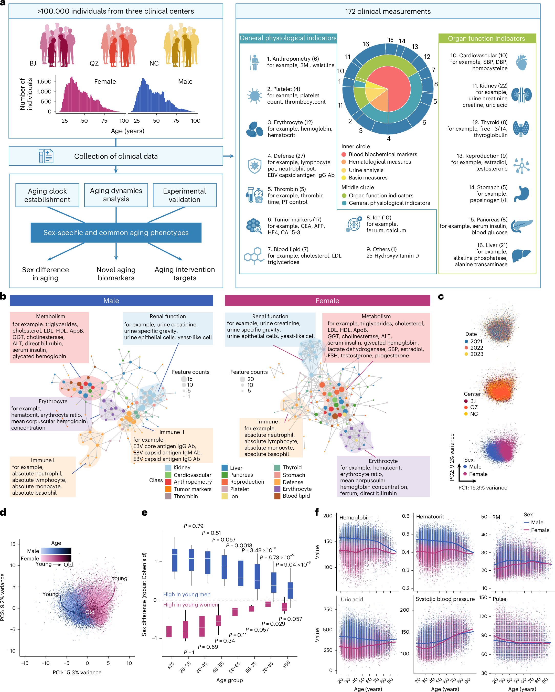
According to the publication record, the work indicates that female and male aging patterns diverge across the organs and molecular layers examined. That framing places the study within a broader shift in aging research, away from a single global score for the whole body and toward measurements that are anatomically and biologically specific.
What a biological aging clock actually measures
The term "aging clock" refers to a computational model trained to estimate a person's biological state from molecular data, then compare that estimate against their chronological age. When the two diverge, researchers interpret the gap as a signal about how quickly or slowly a given tissue or system appears to be aging.
Clocks have been built on several kinds of input. Epigenetic clocks read chemical marks attached to DNA. Transcriptomic clocks read patterns of gene activity. Proteomic clocks read the abundance of circulating or tissue-derived proteins, and metabolomic clocks read small molecules produced by metabolism. Each captures a different slice of biology, and each comes with its own assumptions, noise, and validation requirements.
Why organs, not just the whole body
The title's reference to organs reflects an influential idea in the field: aging is not a single synchronized process. The brain, heart, liver, kidney, skeletal muscle, and immune system can each accumulate damage and functional decline on somewhat independent schedules. A person may show accelerated aging in one organ system while appearing unremarkable in another, and that mismatch is exactly what a whole-body average tends to hide.
The paper's summary does not enumerate which organs were profiled or which clock technologies were used, so the specifics of the panel remain to be read in the full text. What the framing establishes is the ambition: build measurements that resolve aging at the level of individual organs rather than collapsing everything into one figure.

Why sex-specific clocks matter
It is well established that women and men differ in average lifespan and in the timing and frequency of many chronic conditions. Cardiovascular disease, autoimmune disorders, osteoporosis, and several neurodegenerative conditions all show sex-linked patterns in onset or prevalence. Yet many biological aging models have been trained on mixed cohorts and then applied uniformly, which risks masking precisely the differences that drive those patterns.
Sex-specific clocks take the opposite approach. By building and calibrating models separately, or by incorporating sex as a structured variable rather than a nuisance covariate, researchers can ask whether a given molecular signature carries the same meaning in both groups. A marker that predicts decline in one sex may be far less informative in the other, and treating it as universal could produce systematically skewed estimates.
Multi-omics as a cross-check
The phrase "and omics" in the title signals that the analysis is not confined to a single measurement layer. Integrating epigenomic, transcriptomic, proteomic, or metabolomic data offers a way to triangulate. If an organ appears biologically older according to DNA methylation and also according to protein abundance, the finding is more credible than a result resting on one assay alone.
Multi-omics integration also creates its own difficulties. Data layers differ in scale, coverage, cost, and stability. Combining them requires careful normalization and raises the risk of overfitting, particularly when sample sizes are modest relative to the number of features analyzed. Any sex-specific model multiplies that challenge, because splitting a cohort in two reduces the data available for each model.
From measurement to medicine
If organ-level, sex-aware clocks prove reliable, the downstream applications are easy to imagine:
- Risk stratification: identifying which organ system is aging fastest for a given patient, and directing screening toward it.
- Clinical trial design: enrolling participants whose biological, rather than chronological, age best matches the mechanism a therapy targets.
- Intervention monitoring: measuring whether a lifestyle program, drug, or device changes the pace of aging in a specific organ.
- Drug development: using sex-specific signatures to reveal pathways that matter more in one population than another.
- Preventive care: shifting the conversation from calendar age to tissue-level trajectories that can be tracked over time.
None of these uses is automatic. A clock is a statistical construct, and its clinical value depends on whether it predicts outcomes that patients and clinicians actually care about, such as disease onset, functional decline, or mortality, and whether it does so better than simpler measures already in hand.

Caveats worth keeping in view
Several limitations apply to this class of research. Association is not causation: an accelerated clock may reflect an underlying disease process rather than drive it. Cohort composition matters enormously, since age distributions, ancestry, socioeconomic factors, and environmental exposures all shape molecular signatures. Reproducibility across laboratories and platforms remains an active concern. And a fundamental interpretive question lingers, namely how much of any observed sex difference stems from intrinsic biology, how much from hormonal milieu across the lifespan, and how much from social, behavioral, and environmental factors that correlate with sex.
There is also the matter of granularity versus practicality. Organ-level multi-omic profiling can be expensive and, for some tissues, invasive. A clock that requires a biopsy of a specific organ will not scale to routine screening, so translation likely depends on developing blood-based proxies that correlate well with tissue-level signals.
What to watch next
The natural follow-ups are replication in independent and more diverse cohorts, longitudinal tracking to show that clock readings change in ways that predict future health events, and head-to-head comparisons against existing sex-agnostic models. Demonstrating that a sex-specific organ clock adds predictive power, rather than simply adding complexity, is the threshold that separates a methodological advance from a clinical tool.
For now, the paper contributes to a widening recognition that precision medicine has to be precise about sex as well as about genes, tissues, and timing. Aging is not one clock ticking in one place. It is a collection of clocks, and this work argues that they do not run the same way in everyone.
This article is based on reporting by Nature Medicine. Read the original article.

Originally published on nature.com