Routine hospital imaging may be the missing ingredient in medical AI
Artificial intelligence systems for medical imaging are often built from carefully assembled research datasets, but a new study highlighted in Nature Medicine argues that the biggest performance gains may come from something far less curated: the routine scans generated every day inside real health systems.
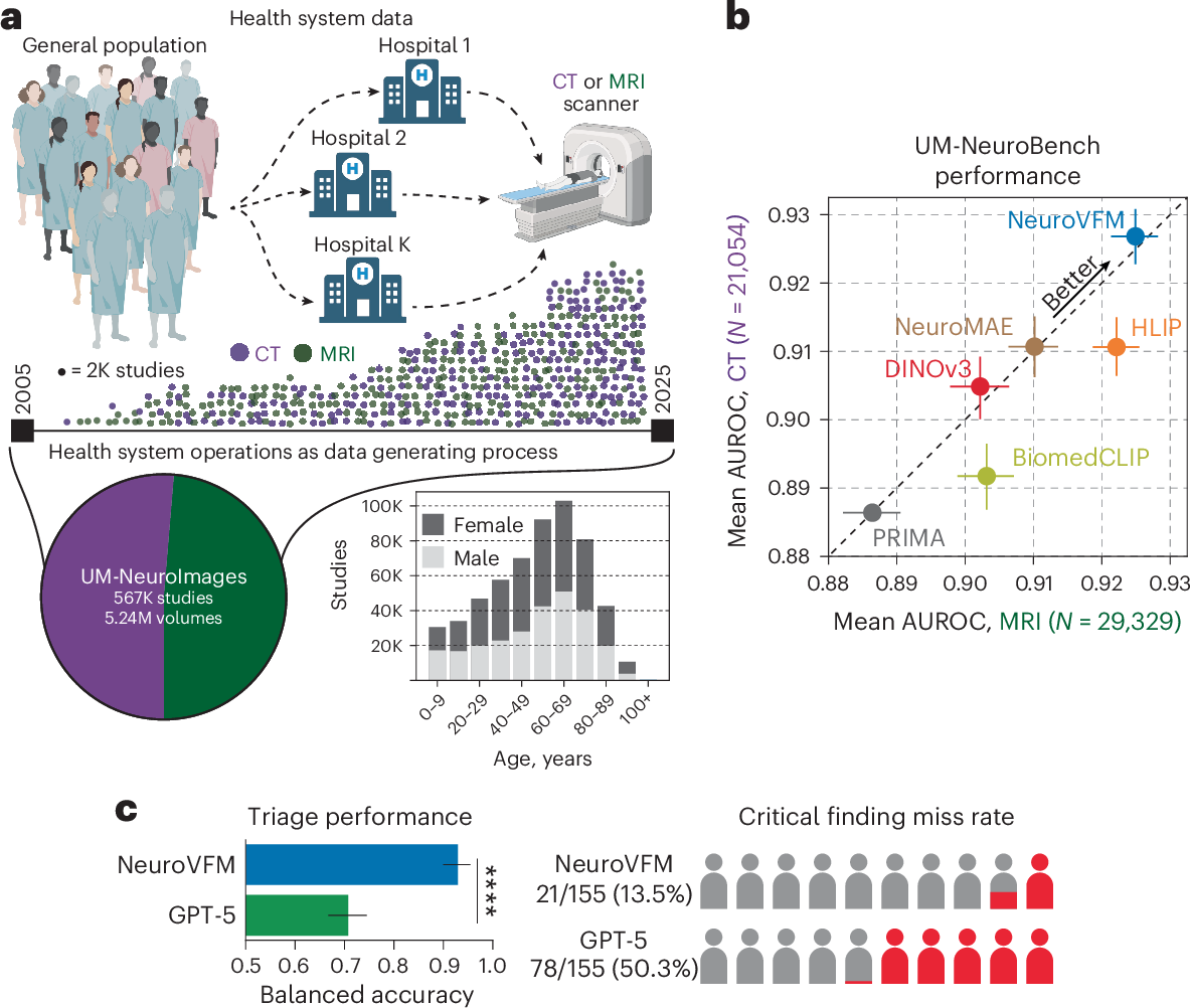
According to the research briefing, investigators trained a three-dimensional visual foundation model on 5.24 million clinical computed tomography and magnetic resonance imaging series. Rather than learning from public internet images or smaller specialist collections, the model was built directly from the flow of everyday neuroimaging used in care. The result, the authors report, was state-of-the-art diagnostic performance along with early signs that the system could support report generation and triage inside real clinical environments.
The work points to a broader shift in medical AI. For several years, the dominant assumption in foundation model development has been that scale alone is the decisive factor and that large public datasets can be adapted to almost any downstream use. This study suggests that for neuroimaging, domain-specific scale matters just as much as raw volume. A model exposed to millions of actual CT and MRI studies appears to learn a shared representation of brain structure and disease that general-purpose training data cannot easily reproduce.
Why real-world data changes the picture
Neuroimaging is a difficult problem for conventional AI pipelines. Brain scans are volumetric rather than flat, they vary across equipment and protocols, and clinically important findings can be subtle, diffuse, or spread across multiple slices. A foundation model trained directly on three-dimensional data from routine practice has a chance to absorb these patterns in a way that smaller supervised datasets often cannot.
The briefing says the model learned a shared representation of neuroanatomy and disease across both CT and MRI. That matters because the two modalities capture different information and are used in different clinical contexts. CT is frequently central in urgent care and acute neurological evaluation, while MRI is often used for more detailed structural assessment. Training across both formats could help produce a model that is more broadly useful across care settings instead of being tied to a single task or scanner type.
The contrast drawn by the paper is notable. The authors report better diagnostic performance than foundation models trained on public internet and medical data. That comparison adds weight to an argument many clinicians and informatics researchers have been making quietly for some time: highly relevant institutional data, if governed and processed correctly, may be more valuable than generic scale borrowed from other domains.
In practice, routine health-system data is messy. It contains variation in acquisition, differences in patient populations, and the artifacts of real clinical workflows. Those properties are usually treated as obstacles. Here, they may have become a strength, because models intended for deployment must eventually function in that same messy environment.

From diagnosis to workflow support
Beyond benchmark performance, the study also points to two practical uses that health systems care about immediately: preliminary report generation and triage. Both remain sensitive applications, but they illustrate where the field is heading.
Preliminary report generation does not mean replacing radiologists. It means an AI system can help summarize probable findings, organize observations, or prepare a draft that a clinician reviews and edits. In high-volume imaging departments, even partial assistance could reduce turnaround times and standardize the first pass through complex cases.
Triage is different but equally consequential. If an imaging AI can identify cases more likely to contain urgent abnormalities, those studies may be surfaced sooner for specialist review. In stroke, hemorrhage, mass effect, or other time-sensitive neurological conditions, minutes can matter. The paper describes triage use as enabled in real health systems, suggesting the model was evaluated with operational deployment in mind rather than as a purely academic exercise.
That combination, strong diagnosis plus workflow assistance, is what makes the study more significant than another accuracy headline. Imaging AI has often struggled to move from laboratory promise to routine use because a model can look impressive on a narrow task but still fail to fit clinical operations. Systems that can support several steps in the chain are more likely to justify the cost and integration effort required for adoption.
What this study does and does not establish
The research briefing provides a concise summary rather than a full operational playbook, so several questions remain open. The excerpt does not specify which diagnostic tasks drove the best results, how performance varied across institutions, or how the model behaved in edge cases. It also describes report generation and triage as preliminary, an important qualifier in a field where early demonstrations can easily be overstated.
Even so, the scale of the training set is difficult to ignore. More than five million CT and MRI series amount to a data resource that only large health systems or multi-institution collaborations can realistically assemble. That raises a strategic issue for the industry. If the most capable medical foundation models depend on health-system archives rather than openly available datasets, the next wave of competition may center on clinical data access, governance, and trust relationships instead of pure model architecture alone.
The study also reinforces a practical lesson for hospitals evaluating AI vendors. Performance claims may depend heavily on what kind of data a model learned from. A tool trained on routine scans collected in care may generalize differently from one adapted from consumer or research imagery. Buyers will likely need sharper questions about provenance, representativeness, and validation before assuming one “foundation model” is interchangeable with another.

A sign of where medical AI is headed
The briefing connects this work to a wider movement in health AI: building general-purpose clinical models from health-system-scale data rather than importing methods wholesale from the public web. That approach is harder because it requires data infrastructure, privacy controls, institutional coordination, and modality-specific engineering. But it may also be the path that produces tools clinicians actually trust.
For neuroimaging, the implication is clear. The future may belong less to models that simply see more images and more to models that learn from the right images: the ones generated in the daily reality of patient care. If those systems continue to show state-of-the-art performance while supporting reporting and triage, they could become foundational not just in name, but in hospital practice.
This article is based on reporting by Nature Medicine. Read the original article.
Originally published on nature.com