A New Layer of Information in Routine Blood Samples
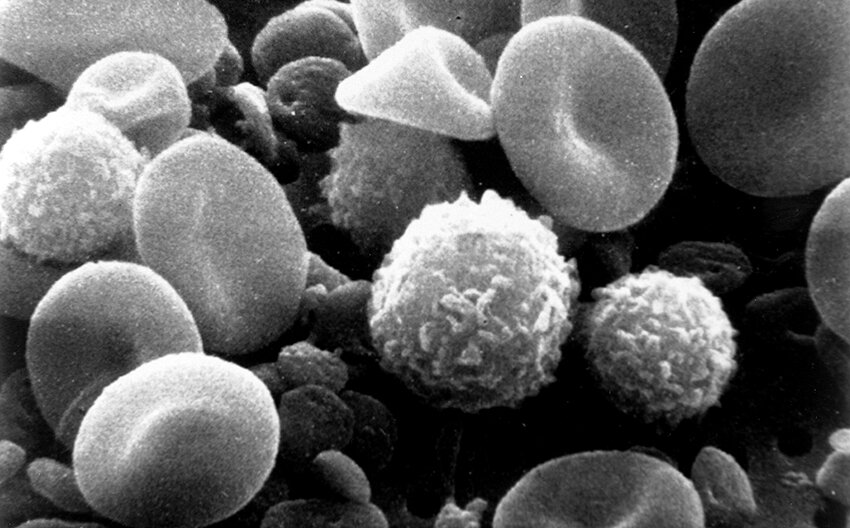
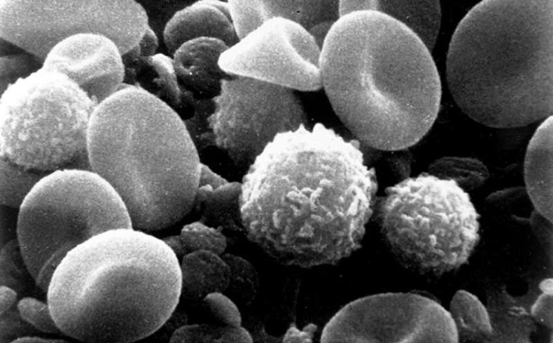
Every routine blood sample contains a diverse population of immune cells, collectively known as peripheral blood mononuclear cells (PBMCs). These cells are a mainstay of clinical and research workflows, offering a window into infections, autoimmune disorders, cancer, and how a patient's immune system responds to therapy. But conventional methods for analyzing PBMCs, which rely on fluorescent labels that bind to surface markers, reveal only which cell types are present—not how those cells are actually functioning. Moreover, the preparation process can alter the cells themselves, potentially skewing results.
Now, a new study published in Biophotonics Discovery demonstrates how advanced optical imaging can expose a previously hidden dimension: the metabolic activity of individual immune cells within a complex, heterogeneous sample. This label-free approach, developed by researchers at the Morgridge Institute for Research and the University of Wisconsin–Madison, could transform how clinicians and scientists interpret the wealth of data contained in a simple blood draw.
Why PBMCs Matter
PBMCs are isolated easily from clinical samples and are already integrated into diagnostic and therapeutic workflows, noted Melissa Skala, senior author of the study and a researcher at the Morgridge Institute and University of Wisconsin–Madison. "PBMCs can be isolated clinically really easily, and they're already used in the clinical workflow," Skala said. "So, the question is, what can we get from them that we aren't already getting?"
That question carries growing clinical weight. PBMCs are routinely studied in conditions as varied as blood cancers, sepsis, lupus, and even cognitive decline. They also serve as the starting material for CAR T-cell therapies, in which a patient's own immune cells are engineered to recognize and attack cancer. In such applications, understanding not just how many immune cells are present, but how metabolically active and fit those cells are, could yield deeper insights into disease progression and treatment outcomes.
For example, a patient with sepsis might have a normal-looking immune cell count, but individual cells could be metabolically exhausted or overactive. Similarly, in CAR T-cell manufacturing, the metabolic health of harvested cells is likely to influence the quality and potency of the final therapeutic product. Yet until now, routine analysis methods have largely overlooked this functional dimension.

Limitations of Conventional Labeling Techniques
Traditionally, measuring immune-cell metabolism has required either isolating specific cell populations or adding fluorescent labels and chemical probes. These strategies come with significant drawbacks. Isolating cells disrupts the natural interplay between different immune cell types, which can be functionally important. Fluorescent dyes and chemical probes can perturb cellular behavior, consume the sample, or provide only a static snapshot rather than a real-time view of cellular activity.
Furthermore, these methods can destroy or permanently alter the cells, making it difficult or impossible to use them for downstream applications like cell therapy. They also tend to treat the sample as a whole, averaging out differences between individual cells that may be biologically significant. A tumor, for instance, might contain immune cells with vastly different metabolic states, and a bulk measurement would obscure that heterogeneity.
The new study sidesteps these issues entirely. It uses a nondestructive technique that measures metabolism in individual immune cells while they remain part of the mixed PBMC population, preserving the natural tissue environment and allowing for further analysis after imaging.
Optical Metabolic Imaging: A Label-Free Approach
The researchers employed a technique called optical metabolic imaging (OMI), which was developed and refined in the Skala Lab. OMI leverages the natural fluorescence of key metabolic coenzymes, specifically NADH and FAD, which are involved in cellular energy production. When immune cells are metabolically active, the concentrations and ratios of these coenzymes shift, and their natural fluorescence changes correspondingly.
By exciting these molecules with low-intensity light and capturing the emitted signals, OMI can map metabolic activity at single-cell resolution without adding any external labels or contrast agents. This allows researchers to observe the living cells in near-physiological conditions and track changes over time.
In the study, the researchers demonstrated that OMI can resolve metabolic differences among immune cell subsets within unmodified PBMC samples. This level of detail has been difficult to achieve with earlier techniques. The method distinguishes between cells that are primarily using glycolysis, a quick but less efficient energy pathway common in activated immune cells, and those relying on oxidative phosphorylation, a more sustainable mode of energy production. These functional distinctions are critical because they correlate with whether an immune cell is quiescent, activated, exhausted, or transitioning into a particular effector state.
Because OMI requires no labeling or physical disruption, it also opens the door to longitudinal studies. The same sample can be imaged repeatedly at different time points, track how immune cells respond to drugs, pathogens, or other stimuli in real time. This is a significant improvement over endpoint assays that only capture a single moment in time.

Clinical and Therapeutic Implications
The findings hold promise across numerous clinical application areas. In cancer immunology, for instance, the metabolic fitness of tumor-infiltrating immune cells is known to influence whether a patient responds to checkpoint inhibitor therapy. If similar metabolic signatures can be detected in PBMCs from a simple blood draw, oncologists might gain a non-invasive way to monitor immune function during treatment.
In CAR T-cell therapy, the process of harvesting, engineering, and expanding a patient's T cells is expensive and labor-intensive. Some CAR T-cell products fail to engraft or lose potency after infusion, which could be linked to poor metabolic condition of the original cells. Using OMI to select the most metabolically robust cells before engineering could improve outcomes.
Autoimmune diseases such as lupus also involve complex immune dysregulation, and PBMCs are often examined to gauge disease activity. A metabolic biomarker derived from OMI could complement current serological markers to give a more comprehensive view of disease state and guide treatment adjustments.
In the context of infectious disease, sepsis continues to be a major clinical challenge. Immune cells from septic patients often exhibit a shift toward an anti-inflammatory, metabolically suppressed state, but measuring this has been difficult. OMI could enable clinicians to accurately assess the functional state of immune cells at the bedside, potentially differentiating between systemic inflammation and immune paralysis in individual patients.
Paving the Way for Clinical Translation
The Skala Lab has been developing OMI for more than a decade and has previously applied it to tissue biopsies, tumor models, and organoids. This newest work extends the approach to blood-based PBMC samples, which are far easier to obtain in clinical settings than tissue specimens.
The fact that PBMCs are already isolated as part of routine clinical workflows is a major advantage. "PBMCs can be isolated clinically really easily, and they're already used in the clinical workflow," reiterated Skala, emphasizing the low barrier to adoption. OMI instruments, while still specialized, are increasingly available in research and clinical laboratories, and the nondestructive nature of the technique means that it could be integrated upstream of existing assays or even cell-processing pipelines.
However, several steps remain before OMI becomes a standard diagnostic tool. Larger clinical studies are needed to validate the metabolic signatures against established disease outcomes and to define what constitutes a "healthy" versus "dysregulated" metabolic profile in specific conditions. The instrumentation must also become more user-friendly and cost-effective for clinical labs. The technical simplicity of label-free imaging, however, is an encouraging sign.
The discovery that routine PBMCs carry hidden metabolic information, accessible with label-free optical imaging, represents a new frontier in immune monitoring. Researchers are now pursuing further studies to catalogue metabolic states across various diseases, and to correlate these states with patient responses to therapy. In the near future, a simple blood sample may offer far more than just a cell count—it may illuminate the functional status of each immune cell within it, guiding personalized treatment in ways that now seem almost futuristic.
This article is based on reporting by Phys.org. Read the original article.

Originally published on phys.org