A new approach to connectomics
Researchers at the University of Illinois Urbana-Champaign have developed a method that aims to make one of neuroscience’s hardest jobs faster and more scalable: mapping how neurons connect to one another. The technique, called Connectome-seq, uses RNA barcodes to label neurons and identify where cells connect at the synapse. The team says the approach captured thousands of neural connections in the mouse brain with single-synapse resolution.
The work, published in Nature Methods, reframes brain mapping as a sequencing challenge rather than a predominantly imaging-based one. That is the core advance claimed by the researchers. Instead of relying mainly on painstaking sectioning, microscopy, and manual reconstruction, the method uses molecular tags that can be read through sequencing workflows.
Why mapping the brain is so difficult
Understanding neural circuits has long been limited by the brute-force complexity of the brain. Traditional mapping methods often require brain tissue to be cut into extremely thin slices, imaged, and then reconstructed piece by piece. That process can be powerful, but it is slow, labor-intensive, and difficult to scale across large numbers of cells and synapses.
Newer sequencing-based methods improved throughput by labeling many neurons at once, but according to the Illinois team, those tools generally reveal where neurons extend rather than which exact cells they connect to at synapses. That distinction matters. A wiring diagram becomes most useful when researchers can identify not just proximity or projection patterns, but actual cell-to-cell communication links.
Boxuan Zhao, a professor of cell and developmental biology at Illinois and the study leader, framed the challenge in engineering terms. If researchers do not know how the brain is wired, he argued, they cannot fully understand its function, optimize their models, or fix what goes wrong when disease disrupts the system.

How Connectome-seq works
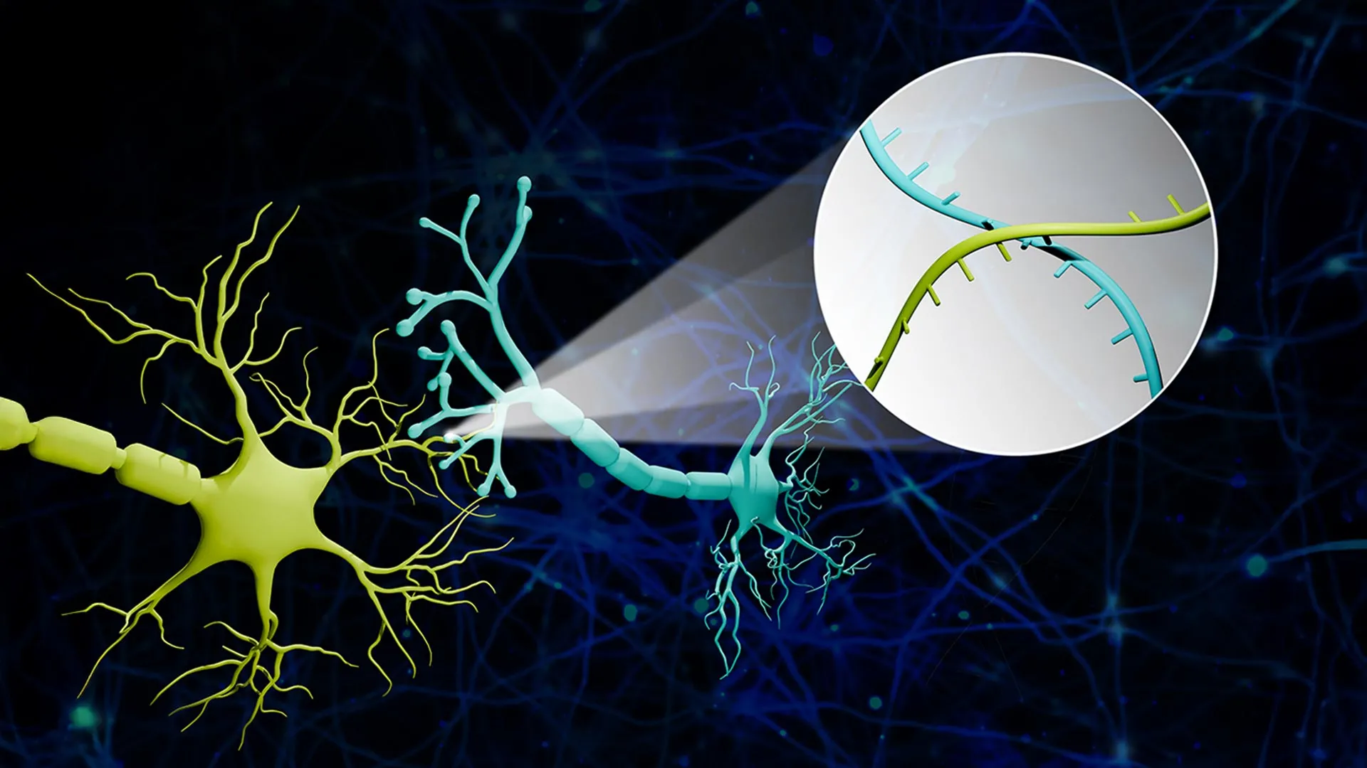
The basic concept behind Connectome-seq is to assign each neuron a unique molecular barcode. Those RNA barcodes then intermingle at the places where neurons connect, allowing researchers to infer synaptic links by reading out paired barcode information. The result is a map of which cells are connected, built from sequencing data rather than from image tracing alone.
In the summary provided by the university, the researchers described the technique as enabling simultaneous mapping of thousands of neural connections with single-synapse resolution. They said this combination of speed, scale, and detail is not available in current technologies.
If that claim holds up through broader adoption, the method could mark an important step for connectomics, the effort to build increasingly precise maps of neural networks. The significance is not only technical. Better connection maps can reveal how circuits are organized, how information moves across networks, and where disease may alter normal architecture.
What the mouse experiments showed
In mice, the method reportedly uncovered previously unknown connections between brain cells. The source summary does not enumerate those specific links, so the clearest supported takeaway is that the team found neural relationships that had not been identified before using existing approaches.
That matters because one of the strongest tests of any mapping method is whether it reveals circuitry that changes how researchers understand a system. A tool that only replicates known architecture can still be valuable, but a tool that exposes new links begins to alter the scientific picture.
The researchers also emphasize that the method works at single-synapse resolution, an important phrase in the field. Synapses are the functional contact points where neurons communicate. Seeing networks at that level increases the chance that a map captures meaningful biological interactions rather than broad anatomical overlap.

Why sequencing could change the pace of circuit science
The deeper implication of Connectome-seq is methodological. Sequencing has repeatedly transformed biology by making once-specialized measurement tasks cheaper, faster, and more scalable. The Illinois team is effectively arguing that a similar transition could happen for circuit mapping if connectivity can be converted into barcode readouts.
That would not eliminate imaging, which remains essential in neuroscience, but it could reduce dependence on the slowest parts of traditional connectomics workflows. A sequencing-friendly approach might let labs examine far more cells, compare many more conditions, and iterate more quickly when studying development, learning, injury, or disease.
It could also make neural circuit analysis more compatible with the rest of modern molecular biology, where sequencing infrastructure and data pipelines are already widely established. The strategic promise is not only better resolution, but better throughput and broader accessibility.
Disease relevance is a major part of the pitch
The Illinois researchers explicitly link the method to neurological disease. Zhao said the technology is directly applicable to understanding circuit dysfunction in neurodegenerative diseases and could provide a platform for circuit-guided therapeutic interventions.
That is an ambitious vision, but the direction is clear. Disorders such as Alzheimer’s and other neurodegenerative conditions are not only problems of cells dying or proteins misfolding. They are also disorders of circuit failure. If researchers can observe how specific connections are lost, rewired, or destabilized, they may be better positioned to identify early changes and design interventions that target affected networks more precisely.
The summary also suggests potential relevance for earlier detection, though it does not claim the method is itself a clinical diagnostic tool. At this stage, the work is a research platform in mice, not a human medical application. The immediate importance is as an enabling technology for basic and translational neuroscience.

What remains to be proven
As with many technical breakthroughs, the next stage is validation and adoption. A new mapping method must show reproducibility, manageable error rates, and usefulness across different brain regions and experimental contexts. Researchers in other labs will want to know how broadly Connectome-seq can be applied, how difficult it is to implement, and how its readouts compare against established methods.
The source material supports a strong claim that the method is faster and more scalable than traditional approaches, but the field will ultimately judge that through repeated use and comparison. Even so, the work fits a larger trend in biology: turning structure into data that can be read, processed, and analyzed at scale.
If Connectome-seq continues to perform as described, it may help move brain mapping away from artisanal reconstruction and toward higher-throughput circuit science. For neuroscience, that would be a meaningful shift. The brain’s wiring has always been central to understanding its function. What has been missing is a practical way to read that wiring at scale. The Illinois team’s answer is to tag the network and sequence the code.
This article is based on reporting by Science Daily. Read the original article.
Originally published on sciencedaily.com