Introduction: A New Lens on Hypertension
Hypertension remains one of the world's leading risk factors for cardiovascular disease, stroke, and kidney failure. Despite decades of research, the molecular underpinnings of blood pressure regulation are still not fully understood, especially how different organs communicate and contribute to the disease. Now, a landmark study published in Science offers an unprecedented view: a cross-organ single-cell analysis that maps the cellular landscape of hypertension across multiple tissues simultaneously. This approach promises to reveal shared mechanisms and potential therapeutic targets that have been invisible to traditional bulk-tissue studies.
The Power of Single-Cell Resolution
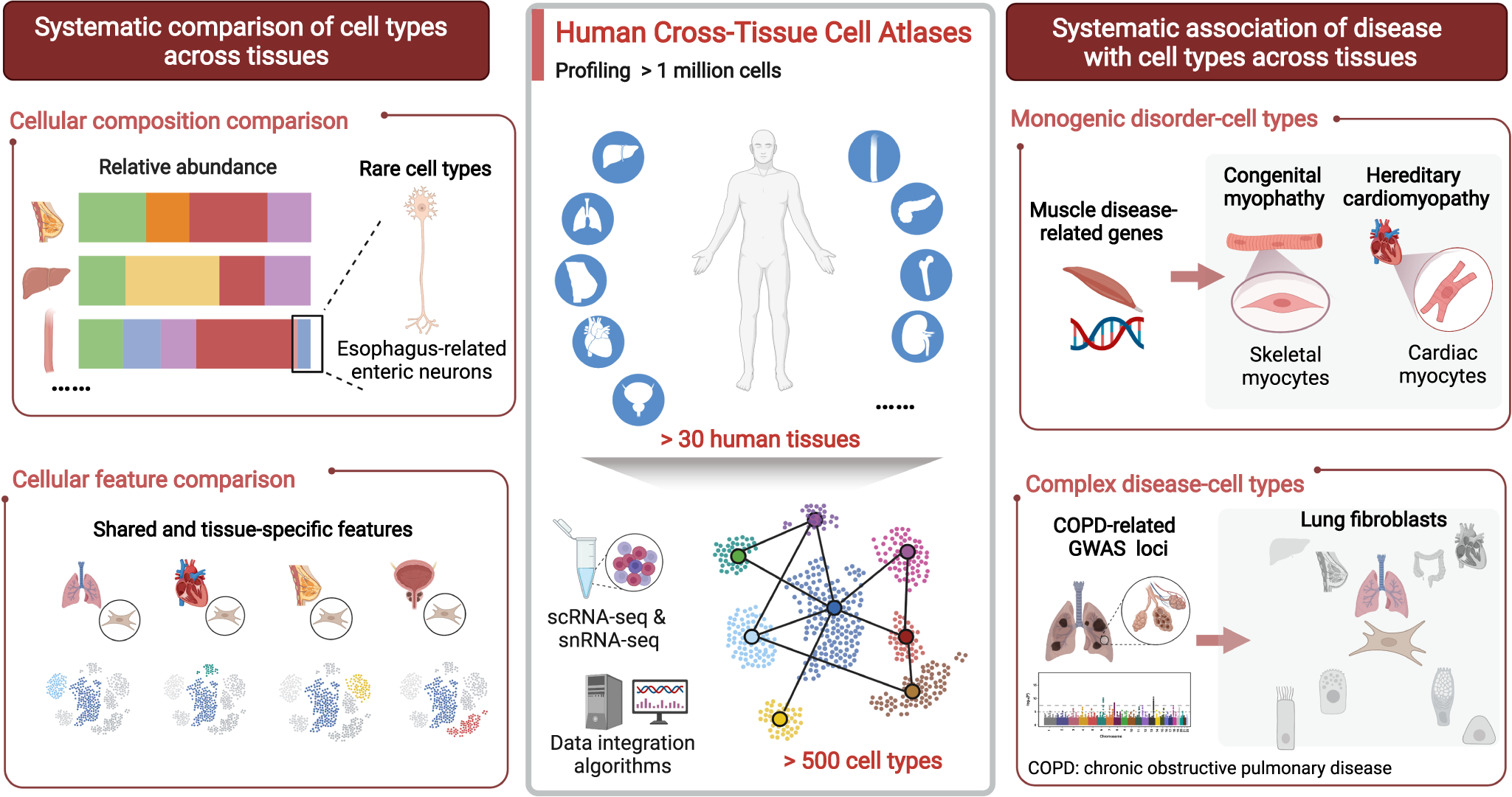
Traditional genomic and proteomic studies of hypertension often rely on homogenized tissue samples, which average out the signals from diverse cell types. This can obscure the contributions of rare but critical cells, such as specific immune subsets or vascular smooth muscle cells in different states. Single-cell RNA sequencing (scRNA-seq) overcomes this limitation by profiling the gene expression of individual cells, allowing researchers to identify distinct cell types, states, and interactions. The new study extends this approach to a multi-organ scale, capturing cells from the heart, kidney, brain, vasculature, and immune organs in hypertensive and normotensive models. By integrating these datasets, the authors constructed a comprehensive cellular atlas of hypertension, revealing how different organs respond and communicate during disease progression.

Shared Immune and Vascular Pathways
One of the most striking findings is the convergence of immune and vascular dysfunction across organs. The analysis identified a common inflammatory signature, with macrophages and T cells showing activated pro-inflammatory states in the kidney, heart, and vasculature of hypertensive subjects. This suggests that systemic inflammation is not merely a consequence but a driver of hypertension, with immune cells migrating between organs and perpetuating damage. In parallel, vascular cells across tissues exhibited signs of endothelial dysfunction and smooth muscle cell remodeling, characterized by altered expression of genes involved in vasoconstriction, extracellular matrix remodeling, and ion transport. These shared pathways highlight potential targets for drugs that could simultaneously address multiple organ systems.
Organ-Specific Insights
While shared mechanisms are important, the study also uncovered organ-specific changes that could inform targeted therapies. In the kidney, the researchers observed significant alterations in tubular epithelial cells, particularly in the proximal tubule, where sodium handling and fluid balance are regulated. This points to a direct link between renal ion transport and blood pressure. In the brain, they found changes in neurons and glial cells within the hypothalamus, a region known to control sympathetic outflow and fluid balance. This suggests that neurogenic mechanisms contribute to hypertension through central pathways. The heart showed signs of early fibrosis and immune infiltration, even before overt hypertrophy, indicating that cardiac damage begins early and may be preventable. These organ-specific insights provide a roadmap for developing precision medicine approaches that consider the unique contributions of each tissue.

Implications for Therapy and Biomarker Development
The identification of novel cell types and gene regulatory networks opens new avenues for therapeutic intervention. For example, the study pinpointed specific ligand-receptor interactions between immune and vascular cells that could be disrupted to reduce inflammation and improve vascular function. Additionally, the authors identified several genes that are consistently dysregulated across organs, which could serve as biomarkers for early detection or monitoring of disease progression. These findings may lead to new classes of antihypertensive drugs that target immune cells or vascular remodeling, rather than the traditional approach of modulating the renin-angiotensin-aldosterone system or calcium channels.
Challenges and Future Directions
While this cross-organ atlas is a major step forward, it also highlights the complexity of hypertension. The study was conducted in animal models, and it remains to be seen how well these findings translate to humans. Future studies will need to validate the identified pathways in patient samples and explore the influence of genetic and environmental factors. Moreover, the sheer volume of data generated by single-cell studies requires sophisticated computational tools to integrate and interpret. The authors have made their dataset publicly available, encouraging other researchers to mine it for additional insights. As single-cell technologies continue to advance, we can expect even more detailed maps that incorporate spatial information and temporal dynamics, bringing us closer to a complete understanding of hypertension.

Conclusion
This cross-organ single-cell analysis represents a paradigm shift in hypertension research. By revealing the coordinated cellular responses across multiple organs, it provides a holistic view of the disease and identifies novel targets for intervention. As we move toward personalized medicine, such comprehensive atlases will be invaluable for tailoring treatments to individual patients based on their unique molecular profiles. The findings not only deepen our fundamental understanding of blood pressure regulation but also offer hope for more effective and targeted therapies in the fight against hypertension.
This article is based on reporting by Science (AAAS). Read the original article.
Originally published on science.org


