Amyotrophic lateral sclerosis, or ALS, has long been understood as a fatal neurodegenerative disease in which the nerve cells that control voluntary movement progressively fail. Immune activity has increasingly become part of the picture, particularly the role of T cells. But a new study suggests that asking whether T-cell activity is simply “good” or “bad” in ALS misses a central issue: where that activity occurs.
Researchers reporting in Nature Medicine found that T-cell responses in cerebrospinal fluid and in the bloodstream were associated with disease progression in markedly different ways. Higher levels of several T-cell populations in cerebrospinal fluid were linked with slower progression, while increases in related circulating T-cell populations were associated with faster progression. The work points to compartmentalized immune dynamics in ALS, rather than a single uniform immune response across the body.
Looking separately at blood and the central nervous system
The researchers combined single-cell RNA sequencing, T-cell receptor sequencing, longitudinal immune profiling and imaging flow cytometry. This allowed them to examine not just broad immune-cell counts, but also the characteristics and persistence of individual T-cell clones in blood and cerebrospinal fluid.
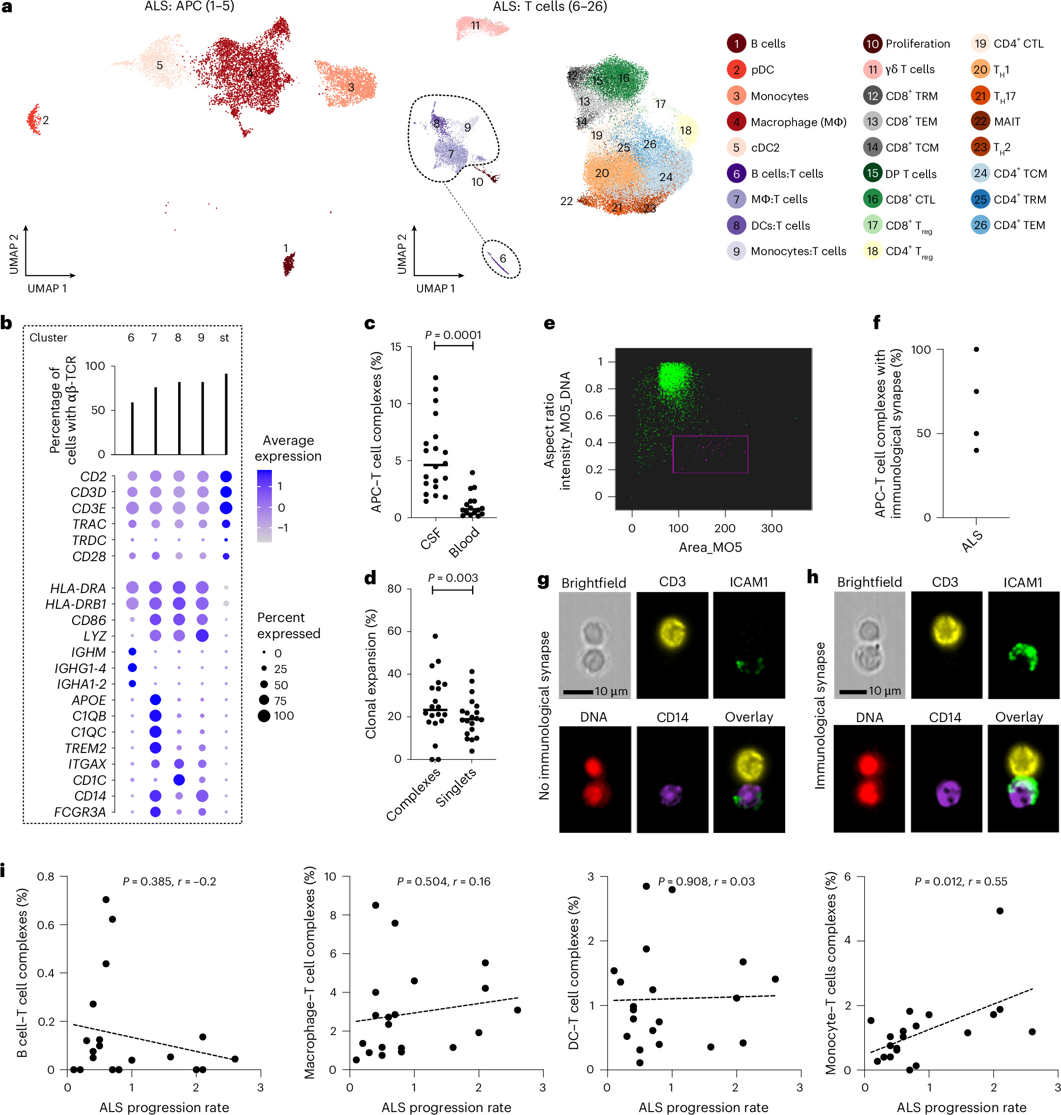
Cerebrospinal fluid surrounds the brain and spinal cord, placing it close to the parts of the nervous system most affected in ALS. The study identified long-lived T-cell clonotypes within this intrathecal, or spinal-fluid, compartment. It also found antigen-presenting cell–T-cell complexes with features consistent with immunological synapse formation, a structure involved when immune cells communicate during antigen presentation.
That observation is important because it supports the idea that immune responses may be actively organized within the central nervous system environment rather than merely reflecting spillover from the bloodstream. The study does not establish a treatment strategy from this finding, but it gives researchers a more specific setting in which to investigate how immune activity may influence the disease.

Associations that run in opposite directions
Among the study’s most consequential findings were the differing links between T-cell frequency and clinical course. Higher frequencies of CD4+ effector T cells, CD4+ regulatory T cells and CD8+ T cells in cerebrospinal fluid were associated with slower disease progression. In contrast, increased circulating CD4+ effector and regulatory T cells were associated with faster progression.
These are associations, not proof that any one cell population directly slows or accelerates ALS. Still, the contrasting pattern offers a potential explanation for why past efforts to interpret immune findings in ALS have been difficult. Measurements drawn from blood alone may not capture the biology occurring near the brain and spinal cord, and similar labels for immune cells do not necessarily mean that those cells are functioning in the same way in different anatomical compartments.
The study also found that T-cell clonotypes shared between blood and cerebrospinal fluid had distinct transcriptional states. In practical terms, genetically related immune-cell populations appearing in both places were not behaving identically at the molecular level. That reinforces the case for treating blood and cerebrospinal fluid as biologically distinct environments when studying ALS immunity.
A viral immune signal linked to poorer outcomes
The researchers additionally examined immune responses related to cytomegalovirus, a common virus that can persist in the body after infection. In people with sporadic ALS, higher frequencies of cytomegalovirus-specific T cells in cerebrospinal fluid, along with anticytomegalovirus antibodies in serum, were correlated with faster progression and shorter survival.
The finding does not show that cytomegalovirus causes ALS or that it is the reason a person’s disease progresses more rapidly. The reported relationship is instead a clue about immune state: virus-specific responses may be part of a pattern associated with poorer outcomes in sporadic ALS. It will require further work to determine whether that pattern reflects a driver of disease, a response to disease processes, or another factor that influences both.

Why the distinction could matter
ALS research needs biological measurements that can help explain differences in how quickly the disease advances and that may eventually guide trials of new therapies. This study suggests that the location of an immune measurement may be as significant as the immune marker itself.
If future research confirms these results, immune profiling could become more informative when it distinguishes systemic activity from activity within the central nervous system. It could also help researchers avoid assuming that an intervention designed to broadly increase or reduce a T-cell response will have the same consequences throughout the body.
The work is especially notable for its longitudinal component. ALS changes over time, and immune activity may change with it. Studying persistent clones and repeated immune profiles gives a more dynamic view than a one-time sample. That is valuable in a disease where the mechanisms linking neurodegeneration, inflammation and clinical decline remain unresolved.
The new findings do not alter the reality that ALS remains a severe and currently fatal disorder. Nor do they identify an established new therapy. But they sharpen an emerging hypothesis: immune responses near the central nervous system may have a different relationship to disease than immune activation in circulation. For a field trying to turn complex biology into clearer targets, that distinction may prove essential.
This article is based on reporting by Nature Medicine. Read the original article.
Originally published on nature.com