CSF testing could add evidence when standard answers are elusive
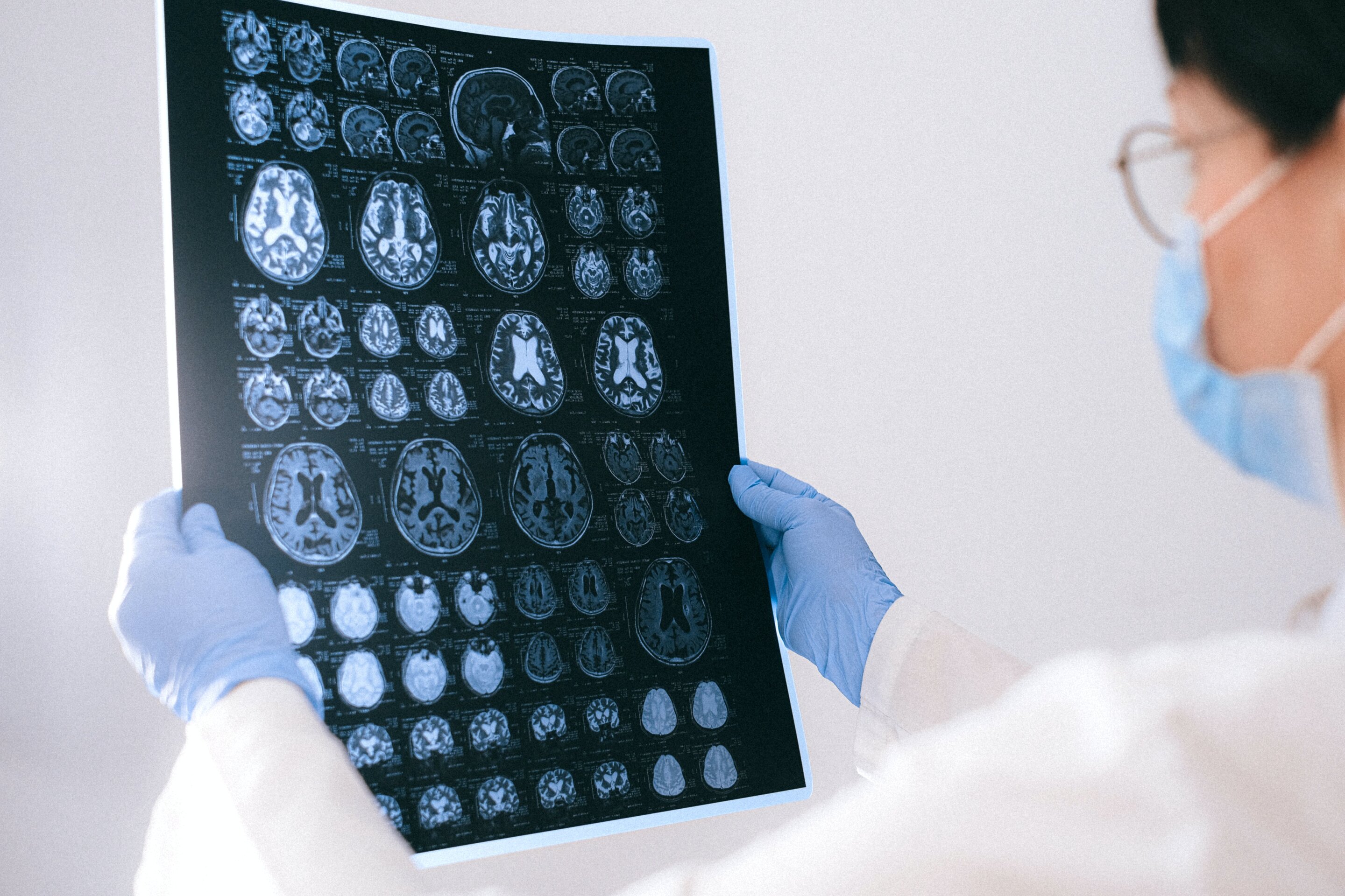
Magnetic resonance imaging and tissue biopsy remain central tools for diagnosing and monitoring cancers involving the brain and spinal cord. But neither can resolve every difficult case. Imaging changes can look alike whether they arise from cancer, inflammation or treatment effects, while a biopsy may be hard to obtain or potentially unsafe because of where a lesion is located.
A Mayo Clinic study published in the Journal of Neuro-Oncology suggests that DNA found in cerebrospinal fluid could provide another piece of information in those situations. The study examined 76 patients facing challenging diagnostic questions involving the brain or spinal cord. Researchers detected tumor-associated DNA in cerebrospinal fluid in 20 patients, or 26% of the group.
The finding does not displace MRI or tissue biopsy. Instead, it points to a possible complementary test that clinicians can weigh alongside scans, pathology and the broader clinical picture when conventional routes do not yield a clear answer.
Why blood tests can be a poor fit for brain tumors
The approach is a form of liquid biopsy: looking for tumor-related material in a body fluid rather than in tissue removed directly from a suspected tumor. Most liquid-biopsy work uses blood, but brain tumors present a particular obstacle. The blood-brain barrier controls what passes between the bloodstream and the brain, which can limit the amount of tumor DNA that reaches blood and make a signal harder to detect.
Cerebrospinal fluid, or CSF, surrounds the brain and spinal cord. Tumor cells can release small fragments of DNA into that fluid. Those fragments, known as cell-free DNA, may carry genetic changes associated with a tumor. CSF can be collected through a lumbar puncture, commonly called a spinal tap.
The appeal is not that the fluid can supply a standalone diagnosis in every case. The Mayo study frames it as an additional source of evidence for cases in which clinicians must distinguish between competing explanations or cannot readily obtain tissue. A positive result may be useful precisely because it can be considered together with other evidence rather than in isolation.

Three clinical situations shaped the study
The 76 patients in the study fell into one of three difficult clinical scenarios:
- An abnormality in the brain or spinal cord without a diagnosis.
- Imaging changes that could represent tumor growth or the effects of treatment.
- Suspected leptomeningeal disease, in which cancer spreads to the membranes and fluid around the brain and spinal cord.
These are settings where a scan may show that something has changed without clearly identifying why. A lesion can also be situated where a tissue sample would be especially challenging or unsafe to collect. In that context, a test that finds tumor-associated DNA in CSF could strengthen the evidence available to a clinical team.
In the study, tumor-associated DNA was detected in one out of roughly every four patients. That proportion also illustrates an important limit: the test did not detect tumor-associated DNA in most of the 76 cases. A negative result therefore cannot simply be read as a universal absence of tumor. The published account instead emphasizes the test as another piece of information for clinicians to interpret with imaging, pathology and the patient’s overall circumstances.
A potential role beside, not instead of, established care
Brain and spinal-cord cancer care often requires decisions under uncertainty. MRI is indispensable for showing anatomical changes and monitoring disease, while tissue biopsy can provide direct diagnostic information. Yet the same imaging appearance can reflect multiple processes, and the risks or feasibility of a biopsy vary by location.
CSF DNA analysis could help narrow uncertainty in selected cases by looking for molecular material in the fluid surrounding the central nervous system. The method is especially notable because it seeks a signal closer to the brain and spinal cord than blood-based liquid biopsies, which may be constrained by the blood-brain barrier.
Still, the results support caution as much as enthusiasm. The study involved 76 patients and reported detection in 20. It describes evidence that can be added to a diagnostic assessment, not a replacement for clinical judgment, MRI or pathology. Whether the result changes management will depend on the particular question facing a patient and on how it aligns with other findings.

What the study adds
The research offers a practical view of where CSF testing may be most useful: not as a routine substitute for established tests, but in the gray areas where those tests leave questions unanswered. That includes ambiguous abnormalities, scans that could indicate either progression or treatment effects, and concern that cancer has reached the leptomeninges.
For clinicians, the value of a molecular signal may lie in combining it with the rest of the case. For patients, the research highlights why a lumbar puncture may sometimes be considered when a scan is inconclusive or obtaining tissue is difficult. It also underscores that results need careful interpretation; a test result is one element of a larger assessment.
Further research will be needed to define how CSF DNA testing performs across tumor types and clinical settings, and how best to incorporate it into decisions. For now, the Mayo Clinic study provides evidence that cerebrospinal fluid can hold useful tumor-related information in some of the brain tumor cases where the usual diagnostic path is least straightforward.
This article is based on reporting by Medical Xpress. Read the original article.
Originally published on medicalxpress.com


