A medical AI designed to show its work
Researchers have reported a clinical vision-language foundation model intended to tackle two persistent limitations of medical artificial intelligence: narrow specialization and opaque outputs. The system, called Explainable Vision Language Foundation Model for Medicine, or EVLF-FM, is described in a study published in Nature Medicine.
Many AI systems used in medical imaging are built for one task, one imaging format or one clinical domain. They may be trained to identify a particular finding in a chest scan, classify a skin lesion or assess an image of the retina. That can produce strong results for a carefully defined problem, but it limits how broadly a model can be used and can make it difficult for clinicians to understand why it reached a conclusion.
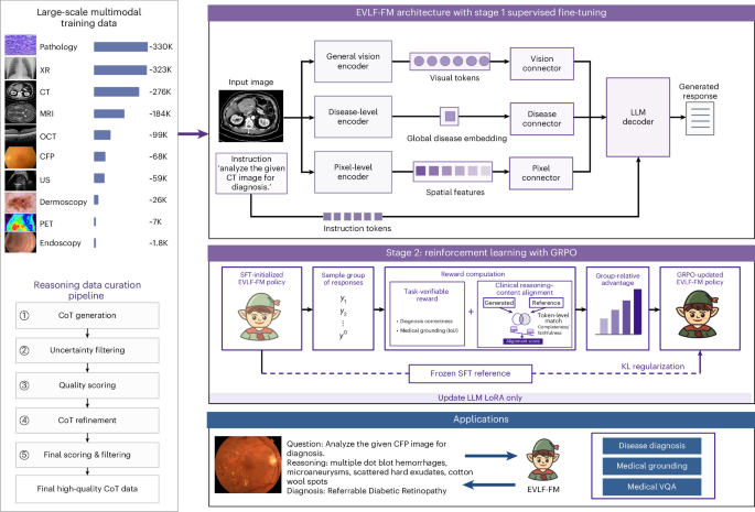
EVLF-FM was developed as a multimodal model: it works with visual clinical data and language-based requests. The research team says it can support disease diagnosis, visual question answering and pixel-level visual grounding. In practical terms, visual grounding is meant to connect an answer to a specific location in an image rather than leaving the user with an unexplained label.
Training across specialties and imaging types
The reported development and testing set included more than 1.3 million samples from 18 global datasets. Those data covered 10 imaging modalities and six specialties: dermatology, hepatology, ophthalmology, pathology, pulmonology and radiology.
That breadth is central to the study’s claim. Medical images vary enormously in appearance, resolution and clinical meaning. A pathology image, for example, is not simply another version of a radiology scan; it has a different scale, visual structure and diagnostic workflow. A model built to operate across such sources has to learn patterns that transfer without treating every specialty as interchangeable.
The authors also evaluated the system externally using 13,443 independent test samples from 17 additional datasets, again spanning 10 imaging modalities. External testing matters because performance on data related to the material used in development can overstate how well a model will fare when it encounters images collected elsewhere.

Reported performance and explainability
For internal disease-diagnostic validation, EVLF-FM achieved an average accuracy of 0.895, which the authors report as the highest among the generalist and specialist models included in their comparison. The paper also reports performance in visual grounding across nine modalities, using accuracy at a 0.5 intersection-over-union threshold.
Intersection over union is a measure commonly used when a model identifies an area in an image. It compares the overlap between the model’s marked region and a reference region. Reporting grounding performance therefore addresses a different question from whether the model selected the right diagnosis: it asks whether the model can point to the relevant visual evidence with reasonable alignment.
The distinction is important for clinical use. A correct-looking answer is not automatically trustworthy, and a highlighted area does not by itself prove a model reasoned in the same way as a clinician. Still, systems that can associate a response with image regions could give users more material to inspect, challenge and compare with their own assessment.
What the study does—and does not—establish
The work presents EVLF-FM as a research model with broad diagnostic and reasoning-oriented capabilities. Its validation across independent datasets is a meaningful part of the evidence, but the supplied study information does not establish routine clinical deployment, regulatory clearance or improved patient outcomes.
Those next steps matter especially for foundation models. A system that works across many tasks may be useful as a common starting point for clinical tools, but it must also be evaluated carefully in the settings where it would be used. Questions include how performance changes across patient populations, devices and health systems; how clinicians respond to the explanations; and whether the model’s outputs improve decisions without introducing new errors or overreliance.
The study nevertheless points toward a different direction for medical AI. Rather than building a separate model for each image type and question, researchers are testing whether a shared system can learn across specialties while making its visual basis more inspectable. If that approach holds up in prospective clinical research, it could help shift medical imaging AI from isolated classifiers toward tools that participate more transparently in clinical workflows.
This article is based on reporting by Nature Medicine. Read the original article.

Originally published on nature.com